Tuesday, May 29, 2012

Lung Cancer and Cigarette Smoke


     I read a paper studying the correlation between rates of cigarette smoking and lung cancer incidence in the United States.  It seems pretty common knowledge today that cigarettes contribute to cancer, but this paper offered some insights into the issue I had previously not thought of.  The paper, found here, analyzes these two things based on different birth cohorts and provides figures showing lung cancer incidence from the early 20th century, lung cancer based on different cohorts and smoking rates in teenagers. However, the figures offer an overly specific analysis with too narrow of a date ranges to show the change in lung cancer incidence in relation to change in smoking rates nationally. In order to get a better visual of how the rates mimic each other I decided to create a figure presenting smoking rates and lung cancer incidence for both males and females in the United States from 1965-2005. The data for smoking came from the CDC and the data for lung cancer came from the American Lung Association.

Here are the figures I used before I combined the lines.

First the lung cancer figure from the American Lung Association:

Transcription Factor KLF11, Diabetes, and Pancreatic Cancer


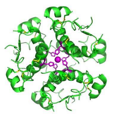
File:InsulinHexamer.jpg
Image Above: Six insulin molecules in a hexamer (6).
KLF11 also known as, TIEG2, is a pancreas transcription factor that has started to gain attention due to its discovered role of being a negative regulator of exocrine cellular growth both found in vitro and in vivo. This is a very interesting case because for the first time KLF11 has been characterized as a glucose regulated transcription factor specific to the insulin gene. In addition, type II diabetes has been linked too two rare variants of the KLF11 gene and has been found to impair transcriptional activity. KLF11 works by altering the TGF-β signaling pathway  binding pathway that can impair the insulin promoter and cause lower levels of insulin release in beta cells.  Two specific diseases that can result in a mutation to the exocrine pathway (which KLF11 is a part of) are pancreatic cancer and type 2 diabetes.   


NovoTFF-100A; A Fancy Way of Saying Brain Tumor Treatment Made Easy

          The new novocure NovoTFF system was officially announced in a press release one year ago May 29, 2012; today, the therapy data collected over the last year will be announced at the annual Scientific Meeting of the American Society of Clinical Oncology (Sacramento Bee).  The system claims to be the future of tumor reduction treatment, and honestly, it does sound like quite the revolution.  The idea behind the system is much more treatment based than "curing" based.  The treatment "contains" the tumor by creating an electrical field at the site, preventing the cancerous cells from dividing, and therefore, the tumor from growing (Ostrovsky).  The draw to the device is the fact that it is supposed to be used throughout the patient's daily life and will not impede their normal activities.  
          Is this the end all cancer treatment program the the Novocure company claims it to be?  Does it merely extend the patient's life a little longer or does it actually allow for tumor and overall cancer reduction?  These are the questions I hope to explore and attempt to answer.

Saturday, May 26, 2012

The Herpes Virus Strikes Again


        The Herpes virus strikes again, though not in the way you might think. In one of my earlier blog (Who Says Herpes Is All Bad), the Herpes Simplex Virus (HSV) was exposed for being more than a menace but actually an innovative tool for marking cancerous cells with Gaussia Luciferase. This marking enables cancer cells that would normally go undetected to glow and be seen. I came across the article, Targeting HSV-1 virions for specific binding to epidermal growth factor receptor-vIII bearing tumor cells, where Paola Grandi and her fellow researchers have found a way to effectively target and destroy deadly cancerous cells through the use of this multi-faced Herpes Simplex Virus (HSV). Grandi found a way to modify the HSV viral envelope glycoprotein’s, by swapping out the normal heparan sulfate binding domain with a tumor-specific immunotoxin. This immunotoxin, not only has the ability to target this modified virus to the naturally found tumor cell biomarkers, but in other research has also shown the capacity to mediate cell death on its own. This type of immunotherapy is a promising alterative to current methods of treatment, especially with their focus on glioblastoma multiform, one of the “most common primary brain tumors [which] are almost universally fatal despite aggressive therapies, including surgery, radiotherapy, and chemotherapy”(1).

Cancer Related Fatigue in Breast Cancer Patients: Exercise As A Therapy to Decrease the Deregulation of Cellular Energetics


Cancer-related fatigue (CRF) is one of the most prominent and under-discussed symptoms of cancer. 48% of cancer patients experience CRF and 58% to 94% of patients with breast cancer experience CRF while they are undergoing treatment (1). The National Comprehensive Cancer Network defines CRF as “a distressing persistent, subjective sense of physical, emotional and/or cognitive tiredness or exhaustion related to cancer or cancer treatment that is not proportional to recent activity and interferes with usual functioning” (2). Due to the persistent sense of tiredness unrelated to activity, patients often describe feeling paralyzed by CRF, as it interferes significantly with their quality of life. What I found fascinating was symptoms of CRF could be present after the cancer has been treated up until death, meaning something about the cancer, treatment, or both, caused a long-lasting, if not permanent change in the patient on a molecular level (1). It is also worth noting that breast cancer patients who received chemotherapy and radiation versus radiation alone were more likely to be fatigued years after treatment, showing the type of therapy most definitely influences the duration and severity of CRF (1).

Pertuzumab: Herceptin Resistance Fix?

About a week ago I came across an article that described a woman who was diagnosed with metastatic breast cancer and was told that unfortunately, she did not have much left to live. She, her family, and friends shared the below video of her sharing her story of her fight against breast cancer and plead to Genentech to allow an early release of the drug Pertuzamab on the basis of compassionate use. Fortunately, the drug was planned to enter the market early June, so Genentech granted her plea.
This sparked my interest in the drug Pertuzamab and lead me to find out why this woman saw the drug as her only hope for survival.
 

Friday, May 25, 2012

Sex Hormone Linked To Thyroid Cancer


I found an interesting review article about how sex hormones might be linked to the development of thyroid cancer. The article is composed of old and recent studies of this hypothesis.The researchers investigated the expression of estrogen receptors α and β on normal and abnormal thyroid tissue and composed tables to determine if estrogen is a contributing factor in tumorigenesis.


Tuesday, May 22, 2012

Cancer: Disease of Death or Disease of Aging

Hello Everyone!
So I recently had an interesting conversation with my Dad on the characteristics of cancer, and we got into an interesting conversation over whether or not cancer is a disease of aging through the lovely discussions we had in class I was able to support the fact that cancer is a disease of aging discussing the accumulation of mutations in cells overtime. I also found a statistic that stated that 50% of diagnosed cancers are of people who are 65 and older and the number is projected to be 70% in 2030.
Now the interesting statistic is that cancer deaths attributes to 13% of deaths worldwide. This does not make it then the disease of death, it just makes it a contributor to death.

But my questions are the following:
1) Wouldn't the fact that cancer is a disease of aging naturally lead it to be the disease of death?
2) What are the reasons for cancer not being the disease of death?

Now my inferences are that cancer is not the disease of death because there are many other diseases that contribute to death, and thus cancer is not the sole contributor therefore not making it the disease of death.
What do you all think? Is cancer a disease of aging or death, or both?
Looking forward to your thoughts! :)

Benzene is a Carcinogen! WHAT?

Hi guys! I feel like every science major in history has at some point heard or talked about benzene. And the common facts are easy; six membered ring, strange stability. Last week in O-Chem we went a bit deeper into what makes benzene tick and what gives it that strange stability. At one point during class, Dr. Carrasco mentioned that benzene was a carcinogen, another pretty common fact. However, when asked what made benzene a carcinogen, Dr. Carrasco answered with "I really don't know exactly why it is a carcinogen."
At that point, my mind screamed out, BLOG POST!

Monday, May 21, 2012

Diagnosing Pancreatic Cancer using a PET Scan


Image Above: Figure A: an anterior view PET scan of torso.  Figure
B: transversal view PET scan of torso.  Figure C: pancreas with the
head of the pancreas almost absorbing all of the tracer.  Figure D:
whole pancreas up taking tracer.  Figure E: Tracer is excreted into
other organs, which cause them to appear on the PET scan (1).  
As of now detection of pancreatic cancer remains as a very difficult task. Pancreatic cancer is often not diagnosed until stage III or IV and the cancer has often already metastasized.  On top of this, most cases of diagnosing pancreatic cancer involve very invasive methods. However, a scan known as positron emission tomography (PET) is a great method for imagery of the body due to its non-invasive nature and can be specified for tissue metabolism (using the tracers) rather than on tissue mass or x- ray absorption. Thus, an increase in glucose metabolism, a hallmark of malignant tumors, can be monitored with a tracer and picked up on a PET scan.  In the case of a pancreas, injecting 2[18F]-fluoro-2-deoxy-D- glucose can be used to detect pancreatic cancer by looking where in the pancreas the highest concentration of the tracer exists (6).    

Friday, May 18, 2012

The Resilience of Melanoma

In class we’ve learned that one of the hallmarks of cancer is resisting cell death. A cancer cell’s resistance to apoptosis and its manipulation of the cell cycle are essentially what make it a cancer cell. In an article titled Apoptosis and Melanoma Chemoresistance, by Maria Soengas and Scott Lowe from the University of Michigan, the specific type of cancer – melanoma, an aggressive skin cancer – is discussed, and its notorious reputation for being resistant to many current chemotherapy mechanisms. Scientists have identified molecules involved with apoptosis, and their alteration in melanoma, and say these are providing new insights into the molecular basis for melanoma chemoresistance. They are now out to develop new strategies to counter these effects and improve in their battle against the disease.

Thursday, May 17, 2012

How Cancer Evolution is Being Used to try to Inhibit Cancer Chemo Resistance and Recurrence


Cancer evolves at a staggering rate. Research is now being done to look into how the mechanism of cancer cells evolve. More recently neoplasms have started to be looked at as their own ecosystem and within this ecosystem different cells that are more and less fit than the others. This new view could help researchers develop drugs that prevent chemoresistance and reoccurrence.
   

Wednesday, May 16, 2012

Cancer: A Consequence of Aging

Introduction & Background
(2)
Dr. Islas often reminds us that cancer is a “reward for living as long as you do.”   Over the weekend, I came across two studies that prove just that.  The first study was conducted by a group of scientists at the Gene Environment Association Studies (GENEVA).  The second study was led by scientists at the National Cancer Institute (NCI).  Both studies found that large chromosomal abnormalities, some of which are correlated with increased risk of cancer, can be detected in a fraction of people without a prior history of cancer.  Sampling from hundreds and thousands of individuals, the scientists also found that alterations in chromosomes increase with age, specifically after the age of 50, and may be associated with an increased risk of cancer. (1)
The GENEVA consortium is sponsored by the National Human Genome Research Institute (NHGRI).  The NCI and the NHGRI are both part of the National Institutes of Health (NIH).
Both studies were published online on May 6, 2012 in Nature Genetics.

Tuesday, May 15, 2012

The Curious Case of Glioblastoma: Back to the Progenitors


Did you ever want to be a super hero? To have the ability to fly, read minds, or travel through time? With much frustration, I realized in my childhood that these powers were not in my metaphorical deck of cards. Unfortunately, the biological mayhem maker, Glioblastoma, has found a way to travel through time. No DeLorean. No Lightning. Just a steady diet of TGFα (Transforming Growth Factor).
Well... sort of through time. Astrocytes are a form of mature glial cells and are part of the brain’s native defense system. In the case of brain injury, they may also be produced from quiescent progenitors and stem cells to fill damaged voids. This is a good thing. This study highlights what is a bad thing and making efficient treatment of Glioblastoma Multiform extremely difficult. Researchers found that prolonged exposure to TGF promotes the conversion of “adult” astrocytes into neural progenitors and then stem cells (like the ones mentioned above). This is called anaplasia, and is a daunting characteristic of Glioblastoma Multiform (GBM, Stage IV glioma). These cells violate a critical feature of healthy brain function: absolute control over cellular replication.

Extending the Life of Glioblastoma Patients: Gene-Modified Therapy

In a recent article posted by the Fred Hutchinson Cancer Research Center, a novel therapy is shown to improve the length of survival of patients with high-risk forms of glioblastoma. After looking at the primary research article, I noticed the researchers used many of the principals associated with personalized and targeted therpies we've recently discussed in class. But first, a little background information on glioblastoma.

According to Maciej Mrugala, the study's lead neuro oncologist, "glioblastoma remains one of the most devastating cancers with a median survival of only 12-15 months" (1). The current treatment strategy is to surgically dissect the tumor if possible, and then to treat the patient with a combination of radiation and chemotherapy. While the treatments are effective at killing the tumor, the side effects are often so severe that dosages must be reduced or treatments completely suspended. This results in less effective treatment and the faster death of the patient. However, it looks as if Adair and colleagues have developed a treatment that circumvents the common problem of toxicity and suppression of blood stem cells (HSCs).

Not all tumor cells are created equal

Introduction & Background
Stefanie Jeffrey, MD (2)
Earlier in the quarter Dr. Islas taught us that three lines of evidence – biochemical, immunological, and cytogenetic – suggested tumors are monoclonal growths.  Recall in a monoclonal growth, a single cell transforms from normal to malignant and subsequently gives rise to a tumor composed of genetically identical cells.  Together, the three experiments, from different fields, provided strong support that cancer arises from a single progenitor cell gone awry.
Alternatively, a tumor can be polyclonal in origin. In this case, multiple cells transform from normal to malignant, leading to a tumor mass composed of genetically distinct sub-populations of cells. (1) (Fig. 1)
Over the weekend, I came across a study conducted by a group of Stanford scientist who have discovered that cancer cells shed by a single tumor into the bloodstream are genetically diverse.  Some cancer cells have turned on genes that make them more adept at lodging themselves in new places, aiding in their ability to metastasize to new organs (2).  Other cancer cells have an entirely different pattern of gene expression.
The senior author of the study is Stefanie Jeffrey, MD, professor of surgery and chief of surgical oncology research at the Stanford University School of Medicine.  The research was published in PLoS ONE on May 7, 2012.

Monday, May 14, 2012

Can an anxious heart lead to and worsen cancer?

Who knew an anxious heart could have a link to cancer?
Over the winter break I browsed through a book entitled, Healing Begins with the Sanctification of the Heart by Dr. Strydom, a Zimbabwean lady who got her training and qualification to be a medical doctor in South Africa. Dr. Strydom, in her insightful book discusses how anxiety and stress can be detrimental to one's health. According to her research, stages 2 & 3 of stress (contributors to prolonged stress) cause excessive production of stress hormones in the human body including ACTH- 'the fear hormone' and cortisol. Cortisol in excessive amounts kills T cells of the immune system, and when this happens T & B cells go out of balance and B cells become overactive and begin to produce excessive antibodies, this then means that the immune system no longer functions efficiently and that the individual in question becomes more susceptible to attack by bacteria and viruses. I would like to venture further and hypothesize that knowing what we previously learnt in lecture about some viruses causing cancer, I believe that anxiety can indirectly lead to cancer via the transformation of viruses and v-oncogenes which are not destroyed by the immune system. Furthermore, we know that one of the hallmarks of cancer involves evading immune system destruction(Hanahan, Weinberg), therefore, in this case, if the immune system is already down or functioning at a less than efficient level, cancer cells may just better thrive in such an environment because a weak immune system won't be able to efficiently ward off viruses and cancer cells.

'Go Ahead, Eat Me...Oh but Wait...Don't Eat Me!' said the Cancer Cell

In a 2010 article from Stanford School of Medicine, research lead by Mark Chao and Ravindra Majet details interesting insight into the machinations of cancer cells. The researchers and their team discovered that many cancer cells actually carry the wellspring of their own ruination, this is in the form of a protein, calreticulin (CRT), an 'eat me' signal on the cell surface of cancer cells that signals circulating immune cells to engulf and digest them. So how come cancer cells are not destroyed efficiently by microphages? Now this is where it starts to get interesting, get this, cancer being the genius that it is also produces separate 'don't eat' me signals in the form of CD47 proteins on cancer cell surfaces creating a process that works to counteract the 'eat me' (CRT) signals.

Previous studies by Stanford scientists involved the characterization of the function of the 'don't eat me' (CD47) protein in cancer cells, through the studies, they established that an anti body that works to block CD47 could be a powerful anti-cancer tool in cancer therapy. They were able to illustrate through their research that anti-CD47 antibodies could eliminate disease in mice transplanted with human myeloid leukemia and also heal a grand proportion of mice with human non-Hodgkins Lymphoma when combined with a second antibody. Although fascinating, the results left the researchers astonished and with a few unanswered questions. This was partly due to the fact that the researchers knew that, CD47 can be found on many cells in the body, yet those cells are unaffected by the CD47 antibody; this observation mystified them. Research also went on to show that normal cell populations do not display CRT and therefore are not expended when they are subjected to CD47 blocking- antibodies.

This research also brought up another question that resonated with me as I was reading this article. That question is whether simply blocking CD47 expression in cancer cells would be sufficient to bring on cancer cell destruction. It turns out that  blocking the CD47- 'don't eat me' cells works to kill cancer cells because cancers like leukemias, lymphomas, and many solid tumors also display CRT-the 'eat me' signal. Interestingly, the most aggressive cancers were found to be the ones producing the most CRT, raising the hope that some of the worst cancers may be the ones most vulnerable to therapies that target CD47 and CRT in particular. This observation also suggests that the immune system works hard to try and get rid of these malignant cancer cells, the high concentration of CRT cells is a testimony to this.

DNA Methylation a Promising Tumor-Specific Marker


In class we have learned that DNA can be changed covalently by the addition of methyl groups to cytosine bases. This alteration is important in shutting down tumor suppressor genes. This article, “DNA methylation in thyroid tumorigenesis” is about how three tumor suppressor genes, CASP8, RASSF1, NIS, are being silenced due to DNA methylation. The clinicians of this study try to identify if maybe methylation is an early change in thyroid tumorigenesis regardless of the cell type. Their main goal is to recognize DNA methylation and that can be used as a biomarker to identify early thyroid cancer.


Avoid an Eye Invasion!


Uveal melanoma is a cancer of the eye involving the iris, ciliary body, or choroid (collectively referred to as the uvea). It is known as melanoma (usually related to skin cancer) because it is still a cancer that arises from the melanocytes, the pigment cells that give color to the skin and the eye.  Uveal cancer cells are known to carry a so called "class 2 signature" which ranks the metastatic cells as one of the more aggressive classes of invasive cells.

Recently, doctors at the Washington University School of Medicine in St. Louis conducted studies that show that drugs known as histone deacetylase (HDAC) inhibitors alter the conformation of the aggressive form of DNA in uveal melanoma, which changes the gene expression, which makes these invasive uveal cancer cells less aggressive. HDAC inhibitors have commonly been used to treat seizures.